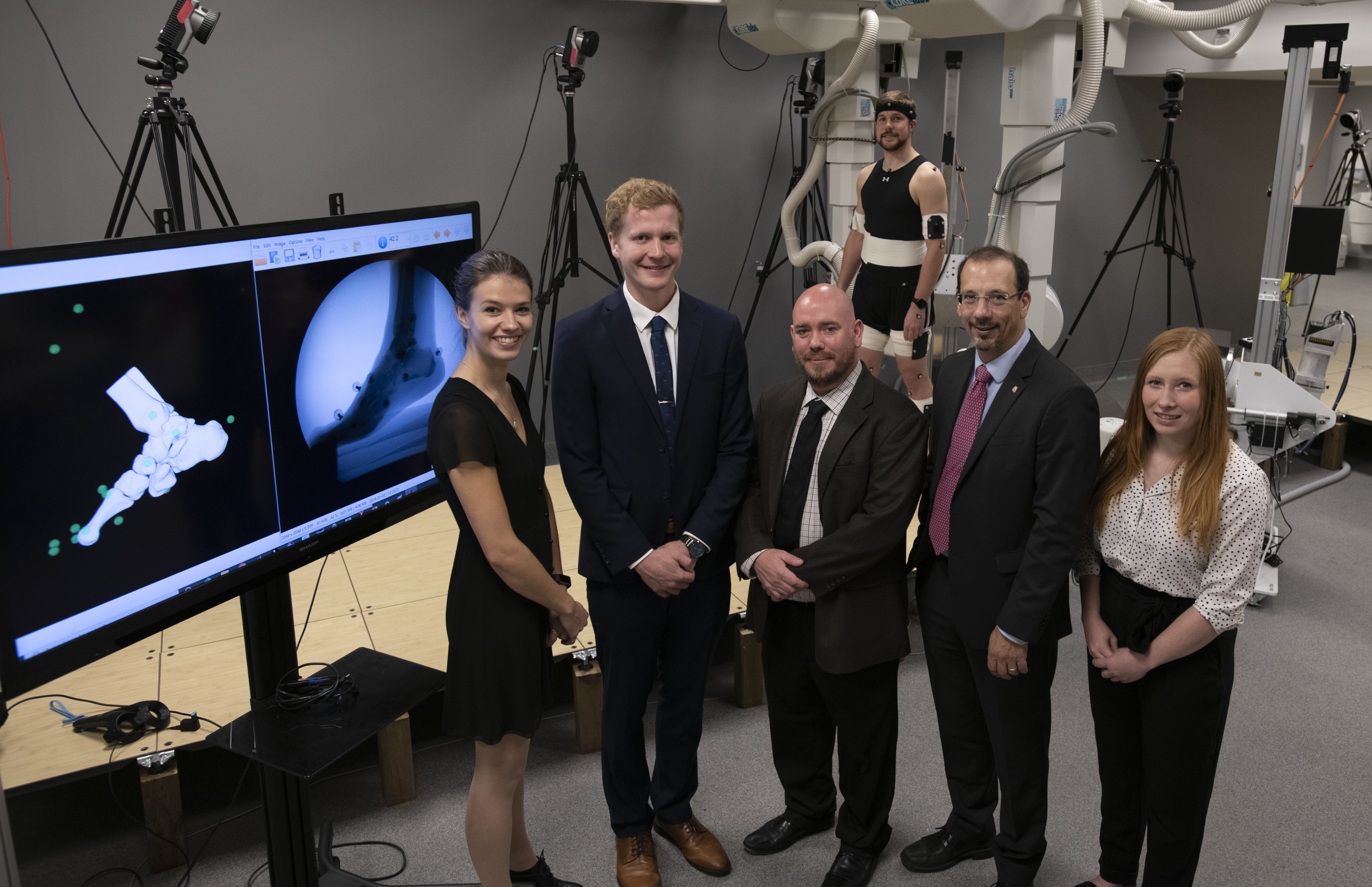
Researcher Dr. Michael Rainbow (centre) and Kevin Deluzio, dean of engineering and applied science, with graduate students (L-R) Lauren Welte, Mitchell Wheatley, Liam Rodgers and Zoe Mack.
Credit
Maclaine Clark
Researchers and doctors at Kingston Health Sciences Centre and Queen’s University are celebrating the completion of a $2.5 million facility that offers unique, “X-ray vision” insights into the biomechanics of nearly any joint in the human body.
While advances in orthopedic medicine have improved the mobility of humans worldwide, the precise workings of our skeleton and its joints are still notoriously difficult to understand, particularly when they’re in motion.
The Skeletal Observation Laboratory, a facility supported by Queen’s University and KHSC at the Hotel Dieu site, is helping to fill that gap. One of only a few such labs in Canada, it offers leading-edge technologies for capturing, in exquisite detail, how the machinery of our bones and joints work when they’re in dynamic action.
Dr. Michael Rainbow, the lab’s lead investigator and Assistant Professor of Mechanical Engineering at Queen’s University, and Dr. David Pichora, orthopedic surgeon and President & CEO of Kingston Health Sciences Centre, demonstrated the marvels of this recently completed lab, including its new, ultra low-dose, load-bearing CAT scanner. Unlike conventional CT machines, which require patients to lie down, with no load on their joints, this scanner can perform 3D scans while the person is standing. It also uses 100 times less radiation dose.
The lab is helping doctors and scientists develop new treatments and preventative strategies tailored to individual bone and joint disorders.
Dr. Rainbow showed how the lab’s powerful imaging equipment -- such as high-speed X-ray, and high speed video capable of 1,000 frames per second -- helps him explore the complex machinery of foot function during walking and running. Better understanding of this complicated network of bones and joints will lead to better designed footwear, prosthetics and orthotics for patients, he says.
Dr. David Pichora talked about collaborating with Dr. Rainbow to study the mechanics of the wrist joint. “The wrist is an extremely complicated joint and a big source of impairment and disease such as arthritis,” he says. “This is a truly world-class lab that helps us to study human motion and the behavior of joints, and design and evaluate treatment options.”
They were joined by graduate students Zoe Mack, who explained her research into wrist mechanics; Mitchel Wheatley, who spoke about the mechanics of the kneecap and why we get knee pain; and Lauren Welte, who spoke on how developing a detailed understanding of healthy foot mechanics can help inform clinical practice.
Later in the day Dr. Rainbow hosted a research talk by Dr. Janet Ronsky, a leading Canadian researcher and innovator in biomechanical engineering and AITF iCORE Strategic Chair in Advanced Diagnostics and Devices at University of Calgary. Dr. Ronsky spoke about how dynamic imaging leads to better understanding of how human joint biomechanics work
The SOL lab is a satellite facility of the Human Mobility Research Centre at KHSC’s KGH site. It is co-located at HDH with the Human Motion Research Lab and the Queen’s Centre for Neurosciences clinical lab, enabling patient-oriented “research from brain to joints.”
Funders of the Skeletal Observation Lab and its research include the Canada Foundation for Innovation (CFI), Canadian Institutes of Health Research (CHIR), Natural Sciences and Engineering Research Council (NSERC), Queen’s Faculty of Engineering and Applied Science and Department of Mechanical and Materials Engineering, The Estate of Donald McGeachy, BSc (Mech Eng) 1940 and University Hospitals Kingston Foundation (UHKF).
Gallery

Researcher Dr. Michael Rainbow (centre) and Kevin Deluzio, dean of engineering and applied science, with graduate students (L-R) Lauren Welte, Mitchell Wheatley, Liam Rodgers and Zoe Mack.


