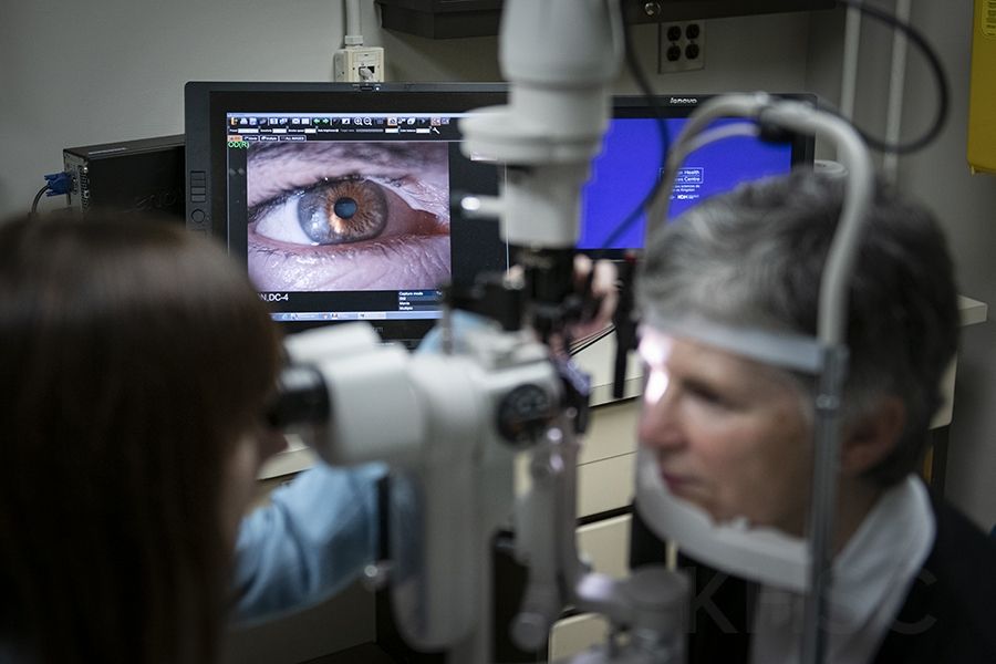
The latest-generation slit lamp packs ramped-up functionality in the form of a high-resolution, digital camera and computer system that precisely images the eye and live streams eye examinations.
Credit
Matthew Manor/KHSC
It’s not often that new technology checks off the big priorities at Kingston Health Sciences Centre all at once but the launch of an advanced digital slit lamp in Ophthalmology is doing exactly that.
In an ocular exam, a slit lamp is vital equipment because it magnifies and illuminates parts of the eye to help detect abnormalities such as cataracts, corneal ulcers or macular degeneration. Typically, a diagnosis results from the use of the slit lamp and physician’s naked eye alone.
But the latest-generation slit lamp now on site at KHSC packs ramped-up functionality in the form of a high-resolution, digital camera and computer system that lets the physician image the eye in precise detail and to live-feed, record and archive eye exams for real-time and future reference.
The technology neatly ticks off KHSC’s priorities of providing quality clinical care, partnering with patients and families and training the health care professionals of the future, says Dr. Martin ten Hove, Head of the Department of Ophthalmology at KHSC and Queen’s University.
“Clinically, slit lamp photography lets us perform a more accurate eye exam,” he says, “because the images pick up minute changes in the eye in a way that’s not possible with the naked eye. That lets us better document complicated eye conditions, such as trauma or corneal ulcers, because we can track the disease process through serial photos. And that leads to earlier diagnosis and treatment.”
The new technology also boosts the partnership between patient and physician by giving patients a real-time look at what’s happening in their eyes.
“The patient who has been diagnosed with an eye infection is more likely to be involved with their eye care when they can actually see pictures of the inflammation and then, at follow-up visits, see images that show how their compliance with treatment is having good results,” says ten Hove. “The technology opens the door to a new level of patient education and teachable moments.”
And it presents huge opportunities from a training and continuing education perspective, says Dr. Rachel Curtis, a third-year resident who has been instrumental in implementing the technology to advance the Competency-Based Medical Education (CBME) component of the Ophthalmology program.
She explains how the new system has capacity for live video streaming that allows clinical preceptors to sit at a computer in one room and observe, in real time, a resident down the hall performing an eye exam.
“Technology like this is central to CBME, where the focus is primarily on coaching and direct observation in order to help learners build their skills and demonstrate competence,” she says.
“Now we have a tool that lets us see and hear junior learners as they connect with patients and perform procedures such as removing a foreign body from an eye. It makes for a very interactive learning environment. And for all learners and faculty, we have a method for documenting and archiving cases. That provides a rich learning and teaching resource for every clinician.”
Three digital slit lamps are now up and running at the HDH site, used mostly in the emergency eye clinic and to help advance the CBME component of the Ophthalmology residency program.
“We originally looked to this technology to boost our CBME resources but then quickly realized how much it contributes to day-to-day clinical practice,” says ten Hove. “Now we would like to spread it across our department, possibly with the help of donor support. We work in a very technology-dependent field and so our sights are always set on obtaining the best equipment possible to improve clinical care.”
Gallery

The latest-generation slit lamp packs ramped-up functionality in the form of a high-resolution, digital camera and computer system that precisely images the eye and live streams eye examinations.


