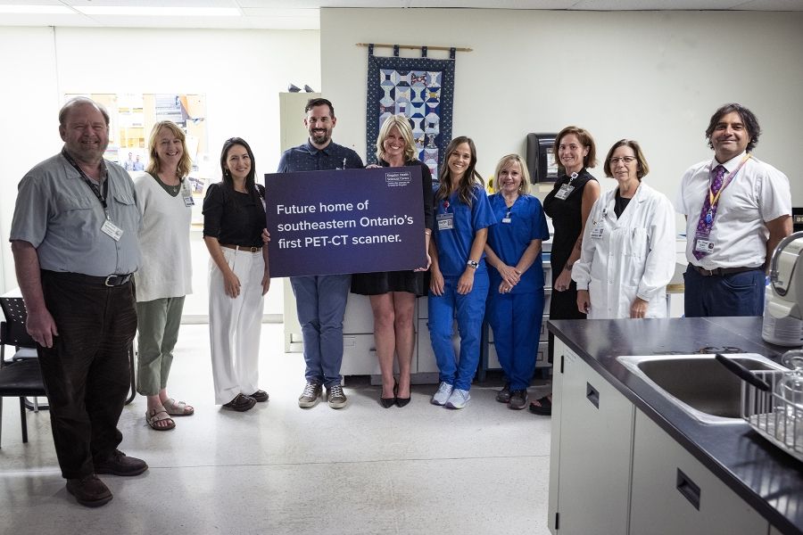
The new positron emission tomography-computerized tomography (PET-CT) scanner at KHSC will combine two types of imaging:
- Positron emission tomography (PET) uses a “tracer” compound to help reveal the metabolic or biochemical function of tissues and organs.
- Computerized tomography (CT) uses specialized x-ray equipment to produce multiple images of the inside of the body.
PET-CT is extremely sensitive and effective for non-invasive detection, diagnosis and for monitoring cancer treatment. It also has applications for cardiac and neurological diseases and will provide advanced imaging for both adults and pediatric patients.
The scanner will mean that patients no long need to travel out of the region to Toronto or Ottawa to receive advanced imaging care.
KHSC’s new scanner
In Sept. 2023, KHSC announced that it would be acquiring Southeastern Ontario’s first PET-CT scanner. This means that patients in the region will soon have improved access to state-of-the-art imaging technology.
Installing a new scanner is a complex and expensive process! Construction is already underway at the KGH site, and is expected to continue for the near future. The total cost for the scanner and associated construction will be approximately $10 million.
The scanner will be installed and hard at work scanning patients by early 2025!
If you’re at the KGH site, you may hear drilling or hammering noises as the foundation is being constructed for the new scanner.
This new device will meet a growing need in our region, and we anticipate we will be able to support approximately 1,000 patients in our first year. PET-CT is a rapidly expanding field. Partnering with GE HealthCare to bring this technology to KHSC, we are thrilled to provide a new service that will have a significant positive impact on patient care in our region. - Dr. Omar Islam, head of Radiology at KHSC
To support the arrival of PET-CT services, KHSC has also recruited a Molecular Imaging Radiologist and will soon be filling the lead PET-CT Technologist position to oversee the planning and implementation of this new service.

